
First of all, let's put light on the meaning of Medical Imaging is the anatomical and physiological visualization and interpretation of the internal structure of the human body.
There are three basic methods in Medical Imaging:
Transmission of energy
Diffusion of energy
Reflection of energy
The methods used in this medical imaging led to the creation of medical imaging systems.

Medical Imaging Systems can be grouped under the following headings:
Radiography Devices
Tomography (CT)
Magnetic Resonance Imaging (MRI)
Nuclear Medicine Devices
Ultrasound
Echocardiography
Radiography Devices use the method of transmitting energy to make imaging with X-ray radiation. For instance, Radiography Devices such as X-ray or X-ray Machines, Angiography, Mammography, Fluoroscopy, C-arm fluoroscopy, and mobile X-ray device can be given as an example. Radiographic Devices are generally divided into two classes:
Conventional Radiography
Fluoroscopy
Conventional Radiography is the filming of the body and organs with X-rays with low doses of X-rays directed only to the area to be examined. With Conventional Radiography, organs such as the lungs and bones can be examined, and lung problems and bone fractures can be detected.

X-ray Machine
With Fluoroscopy, a detailed examination of the wall structure in the organs, blood flow in vessels, and organs in many different areas is provided. The dose of X-ray given in Fluoroscopy is much less than in conventional radiography, but it is continuous. Gastric imaging and Angiography are the most well-known examples. For imaging, auxiliary substances called contrast agents can also be used to better show the functioning of the internal organs. These substances can be barium, iodine, or air, depending on the organ used. Fluoroscopy can also be used as a guide in surgeries. Examples of these are applications such as nailing a bone to the bone or inserting a pacemaker.

Vessel images obtained by angiography


Fluoroscopy Device C-arm Fluoroscopy Device Angiography Device
If we talk about the advantages of radiography devices: The cost is low, and the amount of dose taken is lower than other imaging systems. Disadvantages are that 3-D (dimensional) images cannot be obtained some reactions on the skin, hair loss, and changes in blood can be detected in long-term use due to ionizing radiation.
Just like X-ray devices, tomography uses the method of transmitting energy to obtain a 2-D image from any plane in the body. The image of 2-D slices obtained by tomography is called a sinogram. In Computed Tomography, these sinogram images are combined to create images with more 3-D anatomical details.
X-rays and the detector revolve around the patient while creating a 3-D image in Computed Tomography.


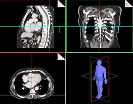
Computed Tomography shows the shape and placement of organs, soft tissues, and bones quite clearly. In addition, CT examinations help doctors in the diagnosis of a simple cyst (formation in the form of a sac with a liquid or semi-liquid substance inside) and a solid tumor (tissue mass formed) to better evaluate the diseases provides. More importantly, CT provides much more detailed images than direct radiography, helping to better visualize the internal structure of organs in assessing the spread of cancers. However, it can cause some problems caused by ionized radiation as an X-ray beam is used.
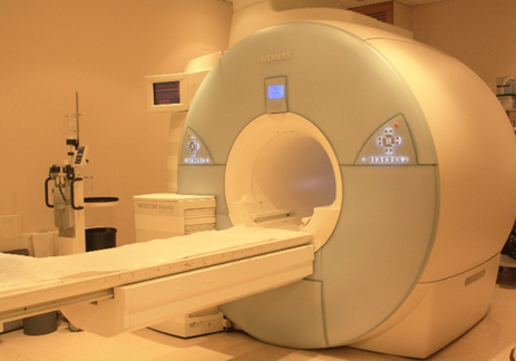
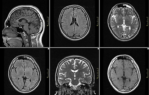
Imaging human soft tissues (organs with high water content) by energy diffusion method is called magnetic imaging. This magnetic imaging is performed by the MRI device with magnets in the device.
During MRI, the powerful scanning magnets in the device align the protons in the same direction, just as the magnet changes the direction of the compass needle. During the shoot, short radio waves are sent to the body at intervals and the protons are out of alignment. When the radio waves are turned off, the protons realign. The movements of the protons during shooting send out radio signals that are detected by the computer.
Protons move at different speeds in different tissues and produce different signals. With the help of a computer, different signals from different tissues are combined on the computer to create a detailed image.


MRI does not contain ionized radiation, so it is safer than other imaging methods. Image creation and creatures in magnets in MRI are achieved by magnetic fields, so they should be used with caution in areas with high metal alloy implants.
MRI is used in all diseases and injuries of the musculoskeletal system, in the diagnosis of cardiovascular diseases, in the detection of all joint problems in the musculoskeletal system, in the diagnosis and treatment planning of ligament and muscle injuries due to sports injuries, in the detection of problems such as tumors, hidden fractures, and osteoporosis, repaired after surgery. It is used for many situations such as monitoring and evaluation of tissues. Again, the diagnosis can be made by applying this method for images such as dementia, nervous system problems, and imaging, which can be encountered especially at an advanced age in the configurations made with the brain.
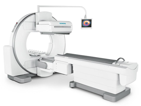
Nuclear Medicine (nuclear medicine-NM) is the use of radioactive products for diagnostic and therapeutic purposes in its own right. Using cameras of different characteristics (PET-CT, SPECT-BT, Gamma cameras, etc.) that actively consume radio substances (radiopharmaceutical drugs) to show the functioning, anatomy, physiology, and pathology of organs such as the heart, transplant, scan, thyroid, expands and brain. is monitored. No radiation is used in Nuclear Medicine. The radioactive substance given to the patient decomposes in the patient's body and provides gamma radiation. Gamma cameras also detect the generated gamma ray and create an image.
With the devices used in Nuclear Medicine, information about the presence and level of the patient such as cancer, dense, vascular organs, metabolic movement, irritations, brain diseases, changes due to aging, and therapeutic operations such as thyroid diseases can be collected.
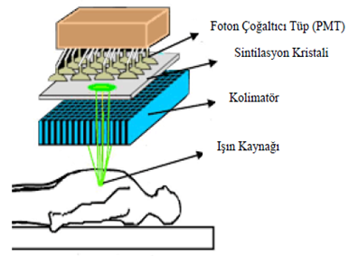
The gamma camera device is a kind of normal camera. It creates an image by detecting gamma rays instead of visible rays reflected from objects. The beam source is the patient body, not the device. Before the imaging procedure at the patient's home, the area to be imaged is used as a special radiopharmaceutical. Technetium-99m, on the other hand, has a half-life of 6 and is the most widely used radioisotope in the Nuclear Medicine imaging department with its 140 keV gamma device.


It is an imaging method formed by the combination of PET (Positron Em Tomography) and CT (Computerized Tomography) devices that enable to obtain of metabolic and anatomical information about PET/CT or PET/CT organs together.


SPECT is a cross-sectional scintigraphy method that enables tomographic imaging of single photon emissions emitted by radiopharmaceuticals given to the body by computer-aided gamma camera systems to display a specific pathophysiological time.


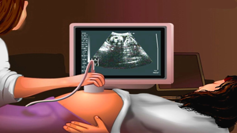
The Ultrasound Device performs simultaneous image tracking by using sound waves at frequencies that cannot be heard by the human ear to move through the tissues. The ultrasound device consists of two separate parts: The first of the parts is the probe that comes into contact with the human body and is controlled manually. The other part of the device is the image processing unit that makes the sound waves sent by the probe visible. So diagnostic ultrasound is a quite safe procedure.
The working logic of ultrasound is that the sound waves are reflected from some parts of the body and detected by the transducer and an image is obtained.
Since the reflection of sound waves from very dense parts of the body (such as bones) or organs with a large air space (such as lungs) will not be at the desired level, imaging such organs with ultrasound is not preferred.
Ultrasound has a wide range of uses in the body:
The uterus and ovaries can be viewed from organs in the genital area, and the baby is followed up during pregnancy. It is effective in diagnosing formations such as stones in the gallbladder. It is used to examine the flow of blood that is not too dense. It can be used with a needle for biopsy procedures or tumor treatment, to determine the exact location, and to make controls organs such as the thyroid.


Trans Esophageal Echocardiography is an endoscopic examination. TEE is also known as cardiac ultrasound or cardiac endoscopy. With a thin tube (probe) lowered into the esophagus through the mouth, the posterior neighborhood of the heart is accessed, and a very clear and detailed image is obtained.

Writer: Arzum Demirbolat / Medical Engineer
Comments